Laptop Color Doppler Ultrasound Scanner Machine Economical Ultrasonic System DW-L3(two probes)
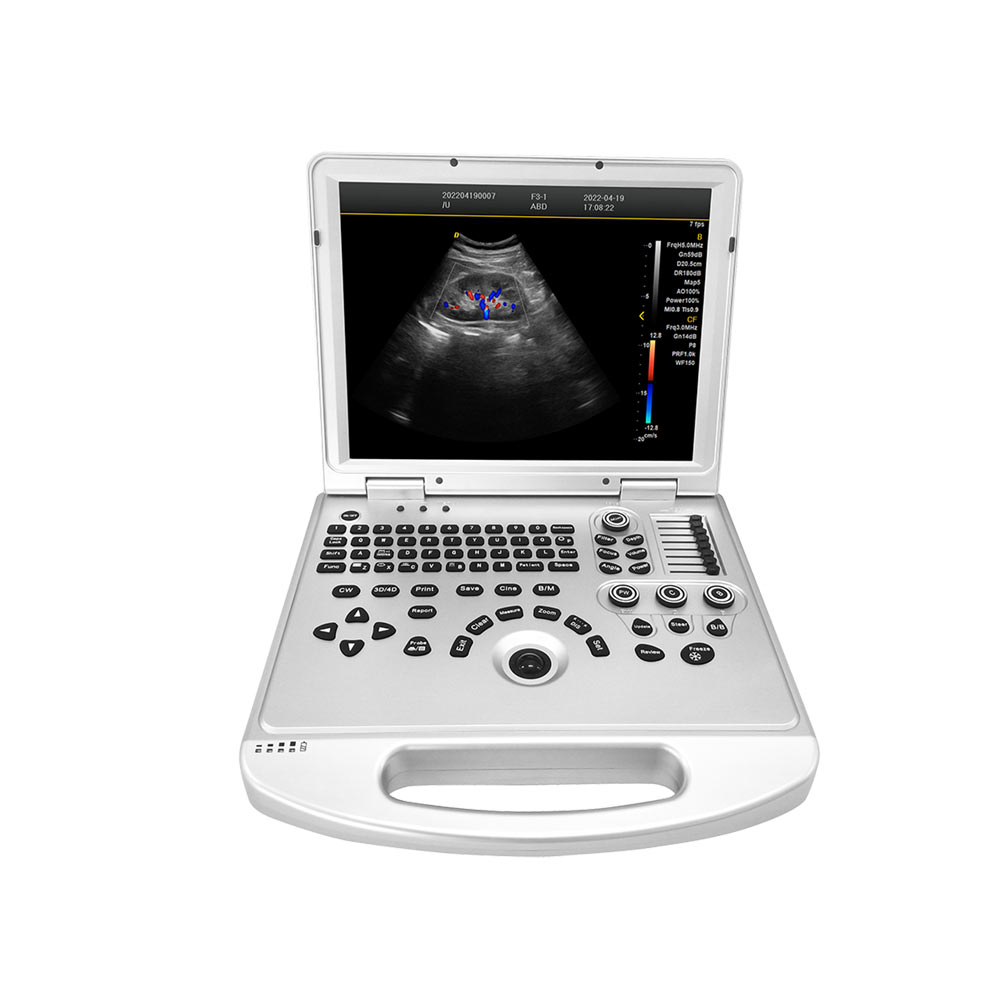
Laptop Color Doppler Ultrasound Scanner Machine Economical Ultrasonic System DW-L3
Specification:
| 1 | Summary of main specifications and system of laptop 4D color Doppler ultrasound |
| 1.1 | Laptop type all digital color Doppler ultrasound host |
| 1.2 | Ultrasonic host operating system: Windows 7 operating system |
| 1.3 | Spectrum pulse-Doppler |
| 1.4 | Direction energy Doppler |
| 1.5 | Real-time three synchronization |
| 1.6 | Space composite imaging: the requirement is 3 level, visual adjustable. |
| 1.7 | Organized harmonic imaging technology |
| 1.8 | 4B imaging mode |
| 1.9 | One key intelligent optimization |
| 1.1 | Support multilingual user interface |
| 1.11 | Monitor: 15 inches, high definition LED |
| 1.12 | Physical clipboard: display the saved image below the screen, and delete it directly. |
| 1.13 | The system has the function of on-the-spot upgrade |
| 1.14 | Presupposition: for different inspections of the viscera, preset the inspection conditions for the best image, reduce the adjustment of the operation, and the commonly used external adjustment and combination regulation. |
| 1.15 | Language: Chinese/English/Russian/Spanish/French/Arabic/Vietnamese/Portuguese/Indonesian |
| 1.16 | The probe interface is 1 |
| 2 | Probes: |
| 2.1 | Convex probe: 2.0MHz/3.0MHz/3.5MHz/4.0MHz/5.5MHz, |
| 2.2 | Linear probe: 6.0MHz/6.5MHz/7.5MHz/10.0MHz/12.0MHz, |
| 2.3 | Trans-vaginal probe: 5.0MHz/6.0MHz/6.5MHz/7.5MHz/9.0MHz, |
| 2.4 | R11 Micro convex probe: 5.5MHz/6.0MHz/6.5MHz/7.5MHz/9.0MHz, |
| 2.5 | R15 Micro convex probe: 5.5MHz/6.0MHz/6.5MHz/7.5MHz/9.0MHz, |
| 2.6 | 4D Volume probe: 2.0MHz/3.0MHz/3.5MHz/4.0MHz/5.5MHz, |
| 2.7 | Under each probe, there is a selection of specialist and viscera mode and rapid entry detection. |
| 3 | Two-dimensional imaging mode |
| 3.1 | Gain: 0-100, step 2 visible adjustable |
| 3.2 | TGC:8 segment adjustable |
| 3.3 | Image optimization: visible and adjustable over 6 levels |
| 3.4 | Dynamic range: 0-270dB 15 level visual adjustable |
| 3.5 | False-color: 7, visible and adjustable |
| 3.6 | Smooth treatment: 8, visible and adjustable |
| 3.7 | Edge enhancement: 8, visible and adjustable |
| 3.8 | Sound power: 0-15, step 7%, visible adjustable |
| 3.9 | Display depth: ≥ 320mm |
| 3.10 | Maximum focus number: 4 focal points, which can be moved throughout the whole process. |
| 3.11 | Scan line density 256 visible tunable |
| 3.12 | Grayscale: 1-16 level visible and adjustable |
| 3.13 | Filtering, 3 kinds |
| 3.14 | Scanning range, 50%-100% |
| 3.15 | Frame correlation, 0-4 level, visible and adjustable |
| 3.16 | The screen has 14 forms of real-time display of voice power, probe frequency, dynamic range, pseudocolor, grayscale and so on. |
| 4 | Color imaging mode |
| 4.1 | Color frequency: ≥5 frequency conversion, visible adjustable |
| 4.2 | Color deflection: equipped with |
| 4.3 | C afterglow 8, visible and adjustable |
| 4.4 | Color map: 7, visible and adjustable |
| 4.5 | Color reversal: adjustable |
| 4.6 | B/C split-screen synchronous display function: equipped with |
| 4.7 | Color baseline: 7, visible and adjustable |
| 4.8 | Color line density: adjustable |
| 5 | Spectrum Doppler mode |
| 5.1 | Sampling volume angle correction: -80 degree to 80 degrees adjustable |
| 5.2 | Sampling volume: 0.5mm-48mm visibility adjustable |
| 5.3 | Frequency:≥ 5, visible and adjustable |
| 5.4 | Baseline: 7 adjustable |
| 5.5 | Smooth: 8 files can be adjusted |
| 5.6 | False-color: 7 kinds of adjustable |
| 5.7 | Maximum display blood flow measurement speed 25m/s, minimum resolvable blood flow measurement speed: 0.1mm/s |
| 6 | Measurement and analysis function: |
| 6.1 | General measurement distance, area, angle, time, slope, heart rate, velocity, acceleration, spectrum tracing, resistance index/pulsatility index, etc. |
| 6.2 | Professional software package: abdomen, volume, ratio, obstetrics and Gynecology, small organs, carotid artery, Urology |
| 7 | Graphic and text management system |
| 7.1 | Host built-in ≥120G solid-state hard disk to start fast and stable |
| 7.2 | Movie playback: 600 frames |
| 7.3 | Internal patient file information management system: can record patient number, name, check number, check date and so on, and can be searched and managed by numbering, checking number, name and so on |
| 7.4 | The type of report is 3 |
| 7.5 | One key fast report graphic and text management |
| 8 | Interface |
| 8.1 | USB interface: 3 |
| 8.2 | Audio interface: 1 |
| 8.3 | HDMI interface: 1 |
| 8.4 | LAN interface: 2 |
| 9 | Technology, after-sales service and other requirements |
| 9.1 | After acceptance, the warranty is free for two years |
| 9.2 | The manufacturer has ISO13485 certification and EU CE certification. |
Packing Details:
Metal case package Size: 62*32*52cm
N.W. 6.7kgs
G.W. 15kgs
No reviews found


